Bone Cross Section Under Microscope : C3 and C4 Plants - BSCI 1510L Literature and Stats Guide ... / The jeol ion beam cross section polisher (cp) is widely used for preparing pristine samples prior to high resolution imaging and elemental analysis with the scanning electron microscope (sem).
Bone Cross Section Under Microscope : C3 and C4 Plants - BSCI 1510L Literature and Stats Guide ... / The jeol ion beam cross section polisher (cp) is widely used for preparing pristine samples prior to high resolution imaging and elemental analysis with the scanning electron microscope (sem).. Jump to navigation jump to search. Trabecular bone found in metaphysis and epiphysis, as seen under microscope. Ladda ned bilder, illustrationer och vektorgrafik med cross section human med hög kvalitet till priser som passar projektets budget perfekt. The nuclear cross section of a nucleus is used to describe the probability that a nuclear reaction will occur. Bones are rigid organs that support and protect various organs of the body, produce red and white blood cells and store minerals.
The jeol ion beam cross section polisher (cp) is widely used for preparing pristine samples prior to high resolution imaging and elemental analysis with the scanning electron microscope (sem). Select the lowest power objective lens. Trabecular bone found in metaphysis and epiphysis, as seen under microscope. Thin section of dinosaur bone. It is placed directly above a specimen.
Cross Section Of Spinal Cord Under The Microscope View ... from thumbs.dreamstime.com Calcein labels as seen under fluorescence microscopy. The differences were significant in anterior. When the light that enters the condenser is polarized by placing a polarizer in the filter holder and a second, crossed polarizer at the image plane. Cross section human skin tissue under microscope view. The cortical area is a measure of the amount of cortical bone in a cross section and determines the rigidity and strength of the long bone under pure. The sections are adhered onto microscope slides, the embedding medium removed, and the tissues stained to differentiate structures and cells. The jeol ion beam cross section polisher (cp) is widely used for preparing pristine samples prior to high resolution imaging and elemental analysis with the scanning electron microscope (sem). They build the entire picture, improve your understanding, consolidate the information and facilitate recall.
It is placed directly above a specimen.
The differences were significant in anterior. Find the perfect under microscope cross section cross stock photo. Cross section human skin tissue under microscope view. Note that the bone matrix is deposited in concentric layers called lamellae. This simply involves placing a section of the bone on the microscope stage and viewing. Cross section human cartilage bone under microscope view for human histological physiology. Bones are rigid organs that support and protect various organs of the body, produce red and white blood cells and store minerals. Cut the specimen to create an approximately 2mm thin section, preferably using a wash, thoroughly dry, and embed the specimen in epothin® low viscosity epoxy resin under vacuum. They build the entire picture, improve your understanding, consolidate the information and facilitate recall. Like most sections of bone, it is strong, but it lacks the rigidity of the diaphysis. The edge of the shielding plate is positioned at the point where the cross section observation is desired, and the specimen is irradiated. These bone cells have long branching arms (d) which lets them communicate with. The finished bone section will be bonded to a microscope slide and so the first step is to grind flat and polish the part of the bone that will be glued to the slide.
They build the entire picture, improve your understanding, consolidate the information and facilitate recall. Be careful pushing it under the clips that the cover slide doesn't move or crack. Bone cross section — stock image. Calcein labels as seen under fluorescence microscopy. Move the stage (the flat ledge the slide sits on) down to its lowest position.
Bone Anatomy | Ask A Biologist from askabiologist.asu.edu Like most sections of bone, it is strong, but it lacks the rigidity of the diaphysis. Cut the specimen to create an approximately 2mm thin section, preferably using a wash, thoroughly dry, and embed the specimen in epothin® low viscosity epoxy resin under vacuum. Thin section of dinosaur bone. This slide showing a cross section of the mammalian trachea (wind pipe) contains examples of several different kinds of tissues. Bone cross section — stock image. If you were to look at it in under a microscope, it would. The lining of the trachea consists of a type of this slide contains a section of dried compact bone. The large dark spots are passages for blood vessels and nerves.
Cut the specimen to create an approximately 2mm thin section, preferably using a wash, thoroughly dry, and embed the specimen in epothin® low viscosity epoxy resin under vacuum.
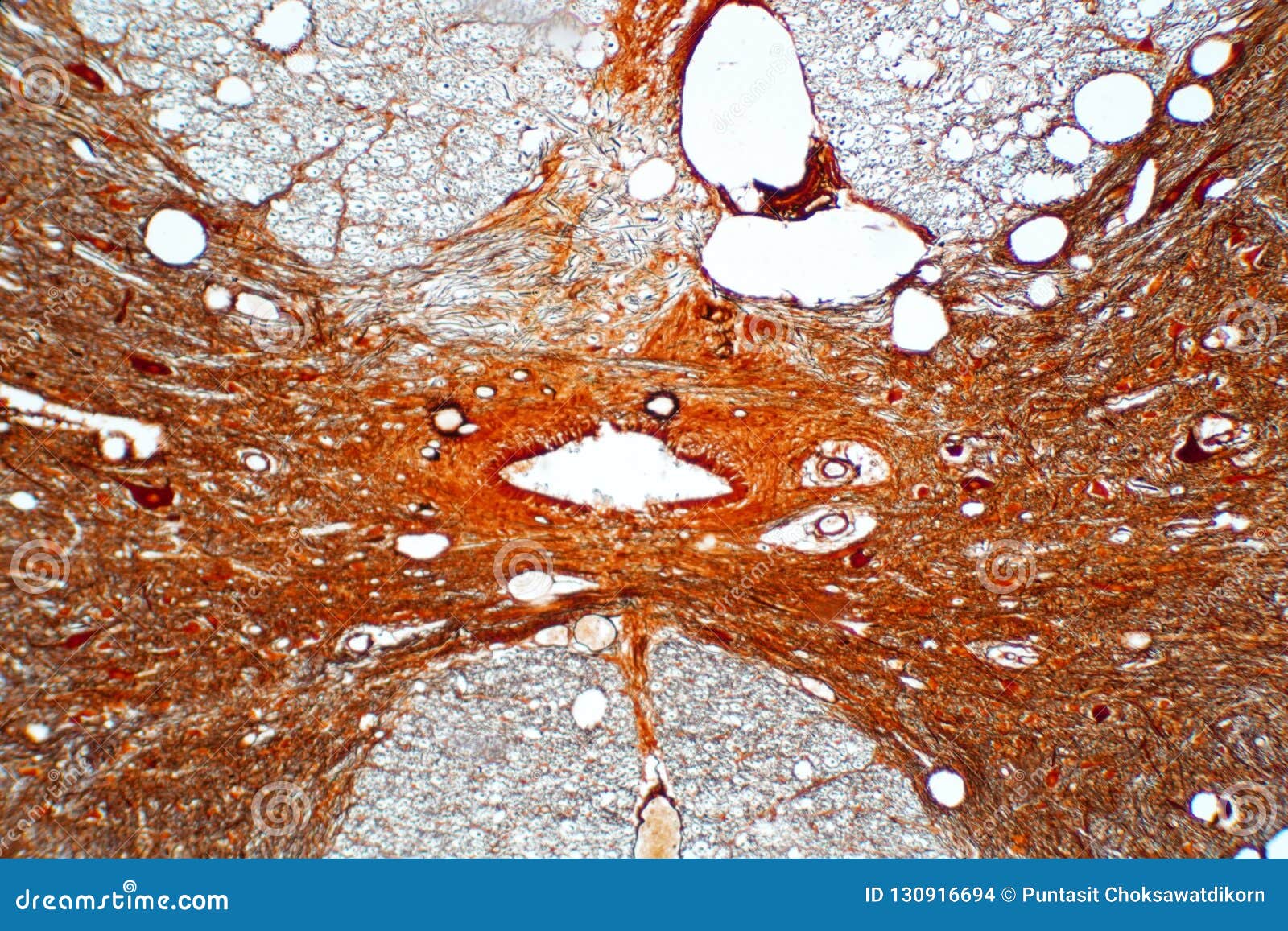
The edge of the shielding plate is positioned at the point where the cross section observation is desired, and the specimen is irradiated. Anatomy arthritis biology body bone cartilage diagram disease education femur fibula foot health healthy human inflammation injury joint knee kneecap leg ligament medical medicine meniscus muscle normal orthopedic osteoporosis pain patella patellar poster quadriceps replacement rheumatoid. This simply involves placing a section of the bone on the microscope stage and viewing. Under the microscope footage of a transverse section of hard bone. The circular patterns are the concentric lamellae of the haversian canal in the center. Note that the bone matrix is deposited in concentric layers called lamellae. Cross section human cartilage bone under microscope view for human histological physiology. Thin section of dinosaur bone. Cross section human skin tissue under microscope view. Like most sections of bone, it is strong, but it lacks the rigidity of the diaphysis. Compact bone cross section courtesy: Ladda ned bilder, illustrationer och vektorgrafik med cross section human med hög kvalitet till priser som passar projektets budget perfekt. To view a bone tissue under the microscope, the bone sample has to be carefully prepared in order to produce a specimen that will provide the best possible results.
Jump to navigation jump to search. The large dark spots are passages for blood vessels and nerves. Like most sections of bone, it is strong, but it lacks the rigidity of the diaphysis. The circular patterns are the concentric lamellae of the haversian canal in the center. The concept of a nuclear cross section can be quantified physically in terms of characteristic area where a larger area means a larger probability of interaction.
Cross Section Human Cartilage Bone Under Microscope View ... from media.istockphoto.com Select the lowest power objective lens. The major components of the cross section polisher (cp) are the ar ion source, shielding plate and specimen, as shown in fig. Like most sections of bone, it is strong, but it lacks the rigidity of the diaphysis. Move the stage (the flat ledge the slide sits on) down to its lowest position. It is placed directly above a specimen. The jeol ion beam cross section polisher (cp) is widely used for preparing pristine samples prior to high resolution imaging and elemental analysis with the scanning electron microscope (sem). Find the perfect under microscope cross section cross stock photo. Be careful pushing it under the clips that the cover slide doesn't move or crack.
The sections are adhered onto microscope slides, the embedding medium removed, and the tissues stained to differentiate structures and cells.
These bone cells have long branching arms (d) which lets them communicate with. In the last decade, considerable technological improvements have been made to repair damaged bones and tissue related posts of bone cross section labeled. Calcein labels as seen under fluorescence microscopy. The large dark spots are passages for blood vessels and nerves. Note that the bone matrix is deposited in concentric layers called lamellae. The edge of the shielding plate is positioned at the point where the cross section observation is desired, and the specimen is irradiated. The concept of a nuclear cross section can be quantified physically in terms of characteristic area where a larger area means a larger probability of interaction. The finished bone section will be bonded to a microscope slide and so the first step is to grind flat and polish the part of the bone that will be glued to the slide. Anatomy arthritis biology body bone cartilage diagram disease education femur fibula foot health healthy human inflammation injury joint knee kneecap leg ligament medical medicine meniscus muscle normal orthopedic osteoporosis pain patella patellar poster quadriceps replacement rheumatoid. Bone marrow is on the upper left. The microscopic cross section measures the probability of occurrence of a particular nuclear reaction. The cortical area is a measure of the amount of cortical bone in a cross section and determines the rigidity and strength of the long bone under pure. Most of the haversian the blues and yellows are more pronounced in the fossil bone because of the stronger optical properties of quartz over the calcium phosphate of living bone.
Compact bone cross section courtesy: bone cross section. Trabecular bone found in metaphysis and epiphysis, as seen under microscope.


0 Comments